
StrokeFlow AI · Inside the workflow

A stroke case, from arrival to treatment.

StrokeFlow AI isn't a feature list. It's a quiet layer inside the acute stroke pathway — one that turns raw CT pixels into a structured, signed report while the clinical team keeps moving. Scroll through a real case.

Research prototype Sub-minute analysis Radiologist-in-the-loop
01The clinical challenge

Every minute costs 1.9 million neurons.

Acute ischemic stroke is a race. Between symptom onset and treatment sit imaging, interpretation, coordination — and a radiologist who may be reading twenty other studies.

StrokeFlow AI exists for one reason: to give that radiologist a calm, structured second read the moment a stroke CT lands in PACS — so the team can decide, faster.

Step 01 of 09
1.9M
neurons lost / minute
14M
years of life lost / hour
< 4.5h
thrombolysis window

Time is brain. The stroke pathway is measured in seconds, not shifts.
02Patient arrives

A code stroke is called.

68-year-old woman, right-sided weakness, last-known-well 47 minutes ago. The ED activates the stroke protocol.

Before StrokeFlow is even involved, the clock is already running. Triage, IV access, NIHSS — the team moves the patient toward CT.

Step 02 of 09
Emergency Department
Suspected acute stroke
Code Stroke
  1. T + 00:00Ambulance arrival
  2. T + 02:14Triage · NIHSS assessment
  3. T + 04:38CT suite ready
  4. 4T + 06:52NCCT acquisition begins
Every minute of delay costs an estimated 1.9 million neurons.
03CT acquisition

Non-contrast CT of the brain.

The technologist runs the acute stroke protocol on the scanner console. Nothing about their workflow changes.

StrokeFlow is invisible at this stage — no new buttons, no separate upload. It listens on the same PACS the department already trusts.

Step 03 of 09
CT · Siemens SOMATOM
Series 3/5 · Axial 1.0mm
Axial brain CT during acquisition
Acquiring · 04.2s remaining
kV 120 · mAs 320 · slice 96/284
W:80 L:40
Modality
NCCT
Study UID
…2f8a·001
Status
Streaming
04Images become available

PACS receives. StrokeFlow receives.

The moment the study is stored in PACS, a copy is routed to StrokeFlow's DICOM listener.

No manual trigger. No exported CD. The examination lands in the AI queue with its priority already set to Code Stroke.

Step 04 of 09
PACS · DICOM Listener
AE Title: STROKEFLOW_AI
Connected
C-STORE received
284 instances · NCCT Brain
0.4s
Study validated
DICOM conformance · patient linkage
0.4s
Routed to AI queue
Priority: Code Stroke
0.4s
Analysis started
Pipeline v2.4 · GPU node 03
Zero clicks. StrokeFlow started before the tech left the console.
05AI-assisted findings

A structured second read, in under a minute.

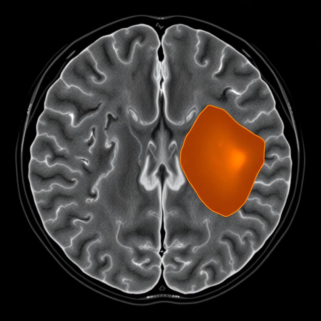
StrokeFlow assesses the study for the four things that matter most at this moment: intracranial hemorrhage, ischemic changes, large vessel occlusion, and ASPECTS.

Findings arrive as a calibrated summary with anatomical overlays — not a black-box percentage. Each result is tied back to the pixels the model looked at.

Step 05 of 09
SF-021 · NCCT · Axial
slice 142 / 284
AI overlay
AI Highlight · Right MCA
AI Findings
Abnormal
ICH
Not DetectedHigh
Ischemia
Right MCAHigh
LVO
Not DetectedModerate
ASPECTS
8 / 10
A quiet layer

StrokeFlow works inside the workflow, not on top of it.

No new console for the technologist. No new portal for the radiologist. The clinical team keeps the tools they trust — StrokeFlow simply makes sure structured information is ready when they open the case.

06Radiologist reviews

The radiologist is always the final voice.

The neuroradiologist opens the case, confirms or adjusts each finding, and signs.

StrokeFlow surfaces evidence, not conclusions. Every AI suggestion is editable; the physician's judgement — and only the physician's judgement — becomes the report.

Step 06 of 09
Dr. J. Smith · Neuroradiology
Reviewing SF-021 · v1 draft
Awaiting Review
Radiologist stays in control. AI is a second reader, never the final voice.
07Structured report

A clean, signed radiology report.

The preliminary AI draft becomes a final, versioned document — with electronic signature, timestamps, and a full audit trail of every change.

Nothing to reformat, retype, or reconcile. The report is ready to leave the reading room the moment the radiologist signs.

Step 07 of 09
Final Radiology Report
v2 · Signed
Patient
SF-021 · 68 F
Study
NCCT Brain · Acute Stroke
Acquired
08:14:22
Signed
08:18:47
Findings

No intracranial hemorrhage. Early ischemic changes in the right MCA territory (insular ribbon, lentiform nucleus). No large vessel occlusion identified on NCCT. ASPECTS 7/10.

Electronic signature
Dr. John Smith, MD
Neuroradiology · Central Hospital
08Case returns to the worklist

Delivered — and visible to the whole team.

The signed report is pushed back into PACS and the EMR. In the StrokeFlow worklist, the case moves from Awaiting review to Signed.

The stroke coordinator, the ED physician, and the interventional team all see the same status, at the same second.

Step 08 of 09
Examinations · Today
Report delivered
SF-021NCCT Brain
Signed · 08:18
SF-016NCCT + CTA
Signed · 07:52
SF-015NCCT Brain
Awaiting review
SF-014NCCT Brain
Signed · 07:14
09Clinical team proceeds

The decision that only humans can make.

With a confirmed read in hand, the stroke team acts: thrombolysis, transfer for thrombectomy, or continued observation.

StrokeFlow's job is done when the clinical decision begins. What remains is measurable: time saved, and — sometimes — brain saved.

Step 09 of 09
Stroke Team Handoff
Neurology · Interventional · ICU
Ready to treat
Door → CT
18 min
CT → Report
6 min
Door → Decision
24 min
Treatment decision: IV thrombolysis initiated. Endovascular team notified for CTA follow-up.
What the workflow adds up to

A quieter reading room. A faster stroke team. A better chance for the patient.

Under one minute

From CT arrival in PACS to a structured AI preliminary report.

Zero new clicks

Runs on the imaging systems and PACS the department already uses.

Radiologist-signed

The final report is always the radiologist's. Fully versioned, fully audited.